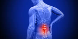
Spinal Stenosis
SPINAL STENOSIS IS OFTEN A SECONDARY CAUSE OF OTEHR OTHER UNDERLYING CONDITIONS: OSTEOARTHRITIS, DISC HERNIATION, DISC DEGENERATION AND TRAUMA
Spinal stenosis is characterized by the narrowing of the spaces within your spine, placing pressure on your spinal cord and nearby nerves. When this occurs, it causes pain, numbness, tingling and weakness. It can develop as the result of a spinal injury or due to thickened ligaments and disc herniation.

NON-SURGICAL TREATMENT Options FOR STENOSIS
Treatment for spinal stenosis depends on the location of the stenosis and the severity of your signs and symptoms.
There are several traditional options that may be a good starting point if the pain is severe and needs to be controlled quickly. Thereafter or if the pain is chronic, mild to moderate there are safer biologic based treatment options.
Traditional options include: Medications, Steroid Injections and Surgery.
More conservative options often combine Physical Therapy, Non-Invasive Laser Therapy and assistive bracing.
The recommendation at PRI is to always start conservative and work your way to surgery being the last option.
To diagnose spinal stenosis, PRI’s medical team will ask you about signs and symptoms, discuss your medical history, and conduct a physical examination. They may order several imaging tests to help pinpoint the cause of your signs and symptoms.
These imaging tests may include:
- X-rays. An X-ray of your back can reveal bony changes, such as bone spurs that may be narrowing the space within the spinal canal.
- Magnetic resonance imaging (MRI). An MRI uses a powerful magnet and radio waves to produce cross-sectional images of your spine. The test can detect damage to your disks and ligaments, as well as the presence of tumors. Most important, it can show where the nerves in the spinal cord are being pressured.
- CT or CT myelogram. If you can’t have an MRI, your doctor may recommend computerized tomography (CT), a test that combines X-ray images taken from many different angles to produce detailed, cross-sectional images of your body. In a CT myelogram, the CT scan is conducted after a contrast dye is injected. The dye outlines the spinal cord and nerves, and it can reveal herniated disks, bone spurs and tumors.
WHAT CAUSES SPINAL STENOSIS?
DISC HERNIATION
Your spine is comprised of a number of bones, also called vertebrae. Each of these bones is cushioned by a spongy disc, which enables spinal flexibility and absorbs shock. When one of these structures bulges or breaks open, it’s called a herniated disc. A herniated disc can occur anywhere along your spine and neck. However, it’s most common in the lower back, or the lumbar spine. We have had great success in treating disc herniation related pain with innovative regenerative treatments like PRP nd BIOLOGICS injections.
DISC DEGENERATION
Disc degeneration and disc tears involve damage to the spinal discs that cushion your vertebrae. While disc tears tend to develop due to a sports injury, age-related degeneration and automobile accidents, disc degeneration typically occurs due to osteoarthritis, a herniated disc or spinal stenosis. In either case, we have achieved great success treating both conditions with our innovative regenerative therapies like PRP and BIOLOGICS injections.
OSTEOARTHRITIS
The most common form of arthritis, osteoarthritis is a degenerative disease characterized by the gradual wearing down of your cartilage over time. Although it can affect any joint in the body, it’s most likely to impact the joints in your spine, hips, knees and hands.
These arthritic changes within the spine cause narrowing of the spine where the nerves exit resulting in spinal stenosis. Osteoarthritis related pain can be successfully treated with PRP and Biologics in place of traditional steroid injections.
Many patients have evidence of spinal stenosis on an MRI or CT scan but may not have symptoms. When they do occur, they often start gradually and worsen over time. Symptoms vary depending on the location of the stenosis and which nerves are affected.
- Numbness or tingling in a hand, arm, foot or leg
- Weakness in a hand, arm, foot or leg
- Problems with walking and balance
- Back and Neck Pain
- Pain or cramping in one or both legs
- In severe cases, bowel or bladder dysfunction (urinary urgency and incontinence)


